


Eye Anatomy: Seeing the Parts that Make Up the Whole
For all the information and insight into vision, eye care, and deep dives into various eye-related concerns or vision-adjacent topics covered in the Optical Expressions blog, it may be we haven’t taken a good look at a detailed anatomy of the eye itself.
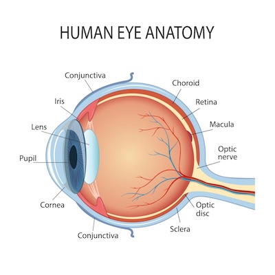
Basic Eye Anatomy Breakdown
- Cornea – The transparent layer forming the front of the eye.
- Sclera – The white outer layer of the eyeball continuous with the cornea.
- Conjunctiva – The mucous membrane that covers the front of the eye and lines the inside of the eyelids.
- Iris – The colored, muscular part of the eye that controls pupil size.
- Pupil – The dark circular opening in the center of the iris, which changes in size to regulate the amount of light reaching the retina.
- Lens – A transparent structure behind the iris that changes shape to focus light onto the retina.
- Retina – A light-sensitive layer of tissue at the back of the inner eye that converts light into neural signals sent to the brain required for vision.
- Optic Nerve – A bundle of over 1 million nerve fibers that transmit visual information from the retina to the brain, facilitating sight.
- Vitreous – A clear, jelly-like substance filling the space between the lens and the retina, which maintains the eyeball's spherical shape, provides structural support, allows light to pass to the retina, and acts as a shock absorber. The vitreous makes up 80% of the eye’s total volume.
- Macula – A highly sensitive area in the center of the retina responsible for sharp, detailed vision.
Peeling Apart the Layers of the Eye
The eye en total is made up of three basic layers.
Fibrous Layer makes up the tough outermost layer of the eye and consists of the sclera and the cornea.
Vascular Layer sits between the fibrous layer and the inner layer, and consists of the iris, ciliary body, and choroid. The choroid is vascular layer (consisting of veins and arteries) between the sclera and the retina that provides nutrients to the retina itself. The ciliary body is a muscular form that connects the iris to the choroid and works to adjust the shape of the lens. The ciliary body also produces aqueous humor, a clear fluid that fills the space between the cornea and the lens of the eye, essential for nourishing eye structures that lack blood supply, maintaining intraocular pressure (IOP), and keeping the eye inflated.
Inner Layer is dominated by the retina, which itself has an outer and inner layer: The outer layer near the choroid and sclera contains the eyes rods and cones, the photoreceptors responsible for converting light into energy. The inner layer of the retina is where the optic nerve is located, taking chemical information from the outer layer and communicating that information to the brain.
The Whole Eye is Greater than the Sum of its Parts
Each part of the eye is a fascinatingly complex and functionally specific component of the whole.
The eye as a complete organ functions as a two-part interconnected system, the anterior (containing the cornea, iris, lens, and aqueous humor) and posterior (containing the vitreous, retina, and optic nerve), which together and instantaneously convert light to chemical signals the brain interprets into the images we see.
Comprehensive Eye Exams from Optical Expressions


Contact Info
Hours of Operation
- Monday 8:00am - 6:00pm
- Tuesday 8:00am - 6:00pm
- Wednesday 8:00am - 6:00pm
- Thursday 8:00am - 6:00pm
- Friday 8:00am - 6:00pm
- Saturday Closed
- Sunday Closed
© 2026 Optical Expressions. All rights Reserved. Accessibility Statement - Privacy Policy - Sitemap
Powered by:
